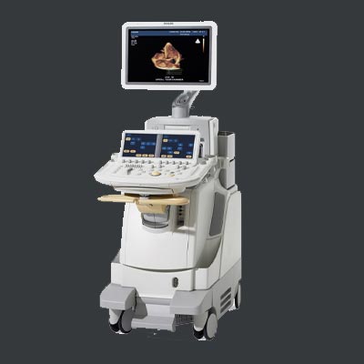
In the procedure of medical imaging and diagnostics, Echocardiography, commonly referred to as ECHO, stands out as a pivotal technique for assessing the heart's structure and function. This non-invasive imaging method utilizes sound waves to create detailed pictures of the heart, offering valuable insights into cardiac health. In this comprehensive guide, we delve into the intricacies of ECHO, exploring how it performs, its diverse applications, and the numerous advantages it brings to the field of healthcare.
What is ECHO?
Echocardiography is a medical imaging technique that employs ultrasound waves to generate real-time images of the heart. By emitting high-frequency sound waves and capturing the echoes as they bounce off cardiac structures, ECHO provides a dynamic visualization of the heart's chambers, valves, and blood flow patterns. This technology has become an indispensable tool for cardiologists and healthcare professionals in diagnosing and monitoring various cardiovascular conditions.

How Does ECHO Perform?
ECHO operates on the principles of ultrasound, a technology that harnesses the reflective properties of sound waves. During an Echocardiogram, a transducer is placed on the patient's chest, emitting ultrasound waves that penetrate the body tissues until they encounter the heart. The echoes produced by these waves are then translated into detailed images by a computer, offering a comprehensive view of the heart's anatomy and functionality. ECHO can capture the heart's movement, identify abnormalities, and assess blood flow dynamics in real-time.
Uses of ECHO in Imaging & Diagnostics
- Structural Assessment: Echocardiography is pivotal in evaluating the heart's structure, detecting abnormalities such as congenital heart defects, valve disorders, and cardiac masses.
- Functional Analysis: It provides real-time assessment of the heart's function, helping diagnose conditions like heart failure and cardiomyopathies.
- Blood Flow Visualization: ECHO enables the visualization of blood flow patterns, aiding in the identification of conditions such as blood clots, stenosis, and regurgitation.
- Monitoring Cardiac Procedures: Echocardiography is used during various cardiac interventions and surgeries to guide procedures and assess outcomes.
- Emergency Diagnosis: It plays a crucial role in emergency situations, helping quickly diagnose acute conditions like myocardial infarction or cardiac tamponade.
Advantages of Echocardiography
- Non-Invasiveness: ECHO is non-invasive, eliminating the need for surgical procedures or catheterization, minimizing patient discomfort and risks.
- Real-Time Imaging: The ability to provide real-time images allows immediate assessment of cardiac function, facilitating prompt decision-making in critical situations.
- Safety: Echocardiography utilizes ultrasound, which is considered safe and does not involve ionizing radiation, making it suitable for repeated examinations.
- Versatility: ECHO can be performed in various settings, including outpatient clinics, emergency rooms, and operating rooms, offering versatility in its application.
- Reduces Cost: Compared to some other imaging techniques, Echocardiography is generally more cost-effective, making it accessible for a broader range of patients.
Echocardiography stands at the forefront of cardiovascular imaging and diagnostics, offering a non-invasive, versatile, and cost-effective approach to assessing the heart's structure and function. Its applications extend across a spectrum of cardiac conditions, making it an invaluable tool in the hands of healthcare professionals striving to enhance patient care and outcomes.
