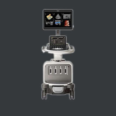
Color Doppler is a pivotal technology in the realm of medical imaging, revolutionizing the way healthcare professionals examine blood flow and diagnose various conditions. This advanced imaging technique utilizes Doppler ultrasound to provide real-time visualizations of blood circulation within the body. In this comprehensive guide, we delve into the intricacies of Color Doppler, exploring how it performs, its diverse applications, and the myriad advantages it offers in the field of diagnostic medicine.
What is Color Doppler?
Color Doppler is a specialized ultrasound technique that extends beyond traditional black and white imaging. It introduces color mapping to represent the direction and speed of blood flow, superimposed onto the grayscale anatomical images. By leveraging the Doppler effect – a change in frequency or wavelength in relation to an observer moving relative to the source – Color Doppler enables the visualization of blood flow dynamics with remarkable clarity.
How Does Color Doppler Perform?
Color Doppler operates by emitting high-frequency sound waves into the body and capturing the reflected signals. The returning echoes are then processed to create a visual representation of blood flow. The color-coded overlay on the anatomical images corresponds to the direction and velocity of the blood, with different colors indicating movement towards or away from the transducer.

Uses of Color Doppler in Medical Imaging
Cardiology
Color Doppler is extensively used in cardiology to assess the blood flow through the heart chambers and vessels. It aids in the diagnosis of conditions such as valvular disorders, congenital heart defects, and coronary artery diseases.
Vascular Imaging
In vascular studies, Color Doppler helps evaluate blood circulation in arteries and veins. It is instrumental in identifying blockages, detecting aneurysms, and assessing the overall vascular health.
Obstetrics and Gynecology
In obstetrics, Color Doppler plays a crucial role in monitoring blood flow in the placenta and fetal vessels. It is employed to identify potential complications during pregnancy, ensuring timely intervention.
Abdominal Imaging
Color Doppler assists in assessing blood flow in organs like the liver, kidneys, and spleen. This is particularly valuable for detecting abnormalities such as tumors or vascular malformations.
Advantages of Color Doppler Imaging
Non-Invasive
Color Doppler is a non-invasive imaging technique, eliminating the need for surgical procedures. This reduces patient discomfort and minimizes the risk of complications.
Real-Time Visualization
The real-time capabilities of Color Doppler provide instant feedback, allowing healthcare professionals to observe dynamic changes in blood flow during the examination.
Enhanced Diagnostic Accuracy
The addition of color mapping enhances diagnostic accuracy by providing a comprehensive view of blood flow patterns, aiding in the identification of abnormalities and precise localization of issues.
Versatility
With applications spanning various medical specialties, Color Doppler is a versatile tool that contributes to comprehensive patient care.
Color Doppler stands as a cornerstone in modern medical imaging, offering a nuanced understanding of blood flow dynamics. Its non-invasive nature, real-time capabilities, and versatility make it an invaluable asset for healthcare professionals across diverse specialties. As technology continues to advance, Color Doppler remains at the forefront, shaping the landscape of diagnostic medicine and contributing to more accurate and timely patient diagnoses.
