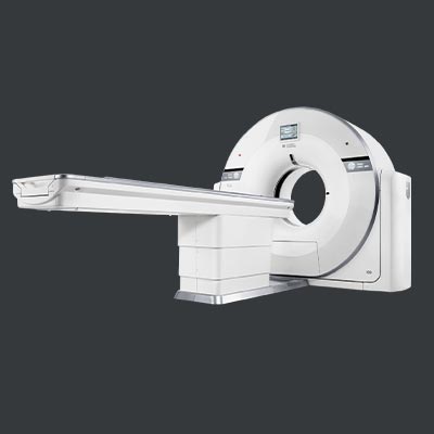
In the realm of medical imaging, the 160-Slice Whole Body & Cardiac CT Scan has emerged as a groundbreaking technology, revolutionizing the way healthcare professionals diagnose and treat various conditions. This advanced imaging technique utilizes a 160-slice computed tomography (CT) scanner to capture detailed images of both the entire body and the heart. In this comprehensive guide, we will delve into the workings of this technology, its performance, key uses, and the advantages it offers in the field of medical diagnostics.
What is a 160-Slice Whole Body & Cardiac CT Scan?
The 160-Slice CT Scan is a cutting-edge medical imaging technique that employs a CT scanner with 160 detector rows to capture cross-sectional images of the body. This technology provides a detailed view of internal structures, allowing healthcare professionals to identify and diagnose a wide range of medical conditions. When applied to cardiac imaging, it specifically focuses on capturing detailed images of the heart and its surrounding structures.
How Does a 160-Slice CT Scan Perform?
The performance of a 160-Slice CT Scan is attributed to its ability to capture a large number of high-resolution images in a single rotation. This enables rapid and precise imaging of the entire body or the cardiac region. The scanner utilizes advanced algorithms to reconstruct these images, creating detailed 3D representations that aid in accurate diagnosis.
Key Features
Cardiac Imaging
- Assessment of coronary artery diseases.
- Evaluation of cardiac structure and function.
- Detection of heart abnormalities such as tumors and congenital defects.
Whole Body Imaging
- Identification and monitoring of tumors.
- Evaluation of vascular conditions.
- Detection of musculoskeletal disorders.
Trauma Evaluation
- Quick assessment of injuries after accidents
- Detection of internal bleeding or organ damag
Advantages of 160-Slice CT Scan
- High Resolution: Provides exceptionally detailed images for accurate diagnosis.
- Speed and Efficiency: Rapid image acquisition allows for quicker diagnosis and reduced patient scan time
- Versatility: Applicable to a wide range of medical conditions and imaging needs.
- Reduced Radiation Dose: Advanced technology allows for lower radiation exposure compared to earlier CT scanners
- 3D Imaging Capability: Enables detailed visualization of anatomical structures in three dimensions
The 160-Slice Whole Body & Cardiac CT Scan stands at the forefront of modern medical imaging, offering unparalleled capabilities in diagnosing and monitoring various health conditions. Its high resolution, speed, and versatility make it an indispensable tool for healthcare professionals, contributing to more accurate and efficient patient care. As technology continues to advance, the 160-Slice CT Scan remains a beacon of innovation in the field of diagnostic imaging.
Services
- CT ABDOMEN PLAIN & CONTRAST
- CT KUB PLAIN
- CT CHEST PLAIN
- CT TEMPORAL PLAIN
- CT CERVICAL SPINE PLAIN
- ct brain with facial bones
- CT ORBITS PLAIN & CONTRAST
- CT SACROILIAC JOINTS PLAIN
- CT PULMONARY ANGIOGRAPHY
- CT AORTOGRAM
- CT FACIAL BONES PLAIN
- CT PERIPHERAL VENOGRAM
- 3D CT JOINT
- CT GUIDED FNAC
- CT NECK PLAIN & CONTRAST
- ct pns full study
- CT CORONARY ANGIOGRAPHY - GRAFT PROTOCOL
- CT VIRTUAL COLONOSCOPY
- CT BRAIN PLAIN
- CT ABDOMEN PLAIN
- CT ENTEROGRAPHY PLAIN & CONTRAST
- HRCT CHEST
- CT ORBITS PLAIN
- CT DORSAL SPINE PLAIN
- CT BRAIN ANGIOGRAPHY
- CT PELVIS PLAIN
- ct tm joints
- CT ABDOMINAL ANGIOGRAPHY
- CT PERIPHERAL ANGIO
- CT DENTAL PLAIN
- CT CISTERNOGRAPHY
- CT NECK VENOGRAM
- CT BRAIN PLAIN & CONTRAST
- CT GUIDED ASPIRATION
- CT CHEST PLAIN & CONTRAST
- CT CORONARY CALCIUM SCORING
- CT VIRTUAL BRONCHOSCOPY
- ct urography
- CT PNS PLAIN
- CT NECK PLAIN
- CT JOINT PLAIN & CONTRAST
- CT JOINT PLAIN
- CT TEMPORAL PLAIN & CONTRAST
- CT LUMBAR SPINE PLAIN
- CT NECK ANGIOGRAPHY
- 3D CT SKULL
- CT RENAL ANGIOGRAPHY
- CT UPPER LIMB ANGIO
- 3D CT PELVIS WITH BOTH HIPS
- CT BRAIN VENOGRAM
- CT PNS PLAIN & CONTRAST
- CT CORONARY ANGIOGRAPHY
- CT KUB PLAIN & CONTRAST
- CT TEMPORAL BONES WITH PNS CUTS

